Laser Surgery to remove Sarcoids

A sarcoid is a benign tumour known as a fibrosarcoma. They can occur anywhere on the horse but are often on the ventral regions as it is believed they are spread from horse to horse by biting flies; so a fly feeds on a horse with a sarcoid and then bites another horse and transmits sarcoid cells to the new host. This hypothesis has been supported by the fact that male horse DNA can be found in female horse sarcoid tissue proving that cells have been transferred from another animal. Another theory is that sarcoids result from infection with bovine papilloma virus but other studies have failed to isolate this virus from equine sarcoid tissue. So the actual cause of sarcoids remains the subject of much debate, but if your horse has one or more sarcoids, what should you do???

Historically, sarcoids have been cut off, frozen with liquid nitrogen, various cytotoxic creams, particularly the “Liverpool” cream produced by Professor Derek Knottenbelt, have been applied and also autologous vaccines have been made from sarcoids in order to stimulate an immune response by the horse to attack sarcoid cells. The “Gold Standard”, particularly for periocular sarcoids, has been to treat the sarcoids with radioactive beads or wires, so a form of radiotherapy. Such treatment has to be done under strictly controlled conditions and is very expensive.

More recently, the use of surgical lasers has become the treatment of choice to remove sarcoids. Diode lasers have been most widely used but they cut slowly and the laser fibre needs constant attention to refresh it during the surgery. At Peasebrook, we have invested in a Carbon Dioxide laser which is much more powerful than the diodes and as you will see from the video below, cuts through the tissue effortlessly. It is essential to take a good margin around the sarcoid and to make sure that all cells in the subsequent crater are also treated. The noise in the video is generated by the smoke evacuation system which is necessary to prevent particles being breathed in by personnel. The wounds can be partially sutured close if required but are generally left to heal by secondary intention. Many lesions can be removed from the standing sedated horse, but if there are extensive sarcoids on the inner thighs, it is safer to do the surgery under general anaesthesia.

The Carbon Dioxide laser is also very efficient at removing facial sarcoids as it is very precise in it’s cutting ability and causes little damage to surrounding tissues. All personnel and the horse, wear protective glasses during the surgery to prevent damage to the eyes.

Sarcoids are a nuisance to the equine world, often causing problems during the vetting procedure. In most cases they can be dealt with and successfully removed, especially if this is done early. Once in a while we come across horses that appear to be immuno-incompetent and succumb to tumour cells, ending up with extensive lesions that reappear whatever treatment is instigated.

Melting Ulcer in a 2 Year old Welsh pony

Melting ulcer as the term suggests, describes a situation where the cornea literally liquifies and changes from a solid structure to more of a gel. The image on the right shows how this case presented at referral.

Melting ulcer on day 1

The cornea as you can see, has stabilised and ruptured has been prevented.

The initiating cause of this condition can be trauma, followed by bacterial infection, viral infection or may result from chemical damage when an irritant inadvertently comes in contact with the cornea.

A careful evaluation of the eye including an ultrasound scan, determined that the innermost layer of the cornea, descemet’s membrane was exposed but not penetrated. Swabs were submitted for bacterial culture and broad spectrum antibiotic cover was instigated. Serum was collected from the pony and used as a topical treatment to provide anti-inflammatory and growth factors to help stabilise the cornea. The eye was lavaged with saline and a mini-tipped swab was used to debride the loose tissue. An in-dwelling subpalpebral lavage system was considered but the patient was very compliant and possible further irritation to the cornea was a risk. The image to the left is two weeks after the start of treatment.

The stabilised cornea is vulnerable to secondary bacterial or fungal infection so preventative medication is still necessary. A further two weeks of treatment were given by which time the healing process was well underway. You will see from the follow-up pictures taken at three weeks and six weeks from the picture above that there is a profound vascularisation from the corneal margins which is essential for healing to take place.

Three weeks after corneal stabilisation

Three weeks after the image above

The eye is now nearly healed but will probably be left with a small scar.

Equine Influenza Vaccination

So why do we vaccinate horses against equine influenza? Clinical signs vary from a mild cough to severe acute respiratory disease but it is unlikely to be fatal. It is however, extremely contagious and can therefore spread rapidly through the equine population. In 2007 an outbreak in Australia, a country in which the vast majority of horses were not vaccinated, resulted in widespread disruption to the equine industry at a cost of around A$1 billion! In the UK, the exact percentage of vaccinated horses is not known, but based on vaccine sales versus the estimated equine population, only 30-40% of horses are covered which is surprisingly low. Equine Influenza is an endemic disease in this country so there are always infected horses about. It is up to us as Veterinary Surgeons to identify potential cases, isolate them and submit samples for testing so that we keep track on this virus. This testing is free due to funding by the Horserace Betting Levy Board and involves taking nasopharyngeal swabs looking for viral DNA by qPCR and paired serum samples looking for an increase in antibody levels. The optimum time to detect via qPCR is between 2 and 5 days following exposure. We are very fortunate in the UK to have the Equine Infectious Disease Surveillance Team (EIDS) funded by the Thoroughbred industry, based at Cambridge University to monitor equine infectious diseases and to advise in the event of an outbreak. The last significant epidemic to occur in the UK was during 2019, when there were 412 confirmed cases on 234 premises. 72% of these cases were unvaccinated. New horses had arrived at the yard within two weeks of a confirmed case in 42% of outbreaks, reaffirming the sense in quarantining/isolating and monitoring clinical signs. Going back to the original question of why we vaccinate; a vaccinated horse will have greater protection in the event of exposure to virus and he or she ,will shed less virus especially if they have had a recent booster. If we want to compete our horses we need to abide strictly to the vaccination requirements of the various governing bodies overseeing competitions. There are some variations between different bodies and we will try and summarise these below: The British Horseracing Authority (BHA) were instrumental in introducing changes to the equine influenza vaccination protocols following the 2019 outbreak.

New equine influenza vaccination rules came into effect on 1 January 2023, following a 12-month transition period to help trainers adjust to the changes.

These require all racehorses to be vaccinated in line with the new primary course and booster vaccination interval schedules set out below: 

As you will see from the table below, some organisations have followed the BHA initiative with respect to primary vaccination courses. It would make everyone’s lives easier if there was one protocol across all disciplines :

Bilateral Iliac Thrombosis in a Thoroughbred Racehorse

Hind limb lameness is a relatively common presentation in racing and sports horses. As we know, there are many possible causes; joint disease often involving OCD lesions, leading to osteoarthritis, soft tissue injury such as proximal suspensory desmitis, and stress fractures typically involving the pastern, cannon bone, tibia or pelvis. All these conditions are relatively straightforward to locate and diagnose. This blog, however, describes a more unusual cause of a transient hind limb lameness, which is brought on by strenuous exercise and then appears to resolve. The video clip below, shows a typical presentation of iliac thrombosis in which a section of the iliac artery, a terminal branch of the aorta supplying the hind leg, becomes partially blocked. The reduced capability of the artery to supply oxygenated blood to the hind limb results in pain and discomfort often causing the horse to stamp or carry the leg. Eventually the oxygen debt caused by the strenuous exercise is resolved as the horse cools down and the pain subsides. The exact cause of the thrombus is unknown; it used to be linked to migrating strongyle larvae but these parasites have been largely eradicated and the condition still occurs. Some other vascular disease must be responsible. Treatment with blood thinning drugs such as aspirin is often attempted but is usually unsuccessful.

Transient hind limb lameness post exerecise

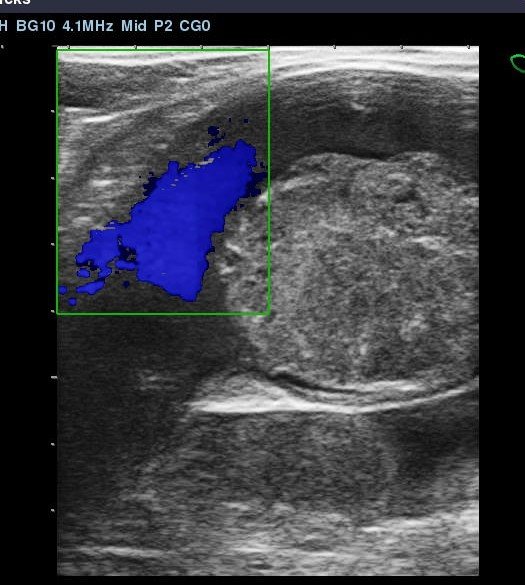
Diagnosis of this condition was confirmed by scanning the iliac arteries using transrectal ultrasound imaging. Normally the arteries are wide and uniform in cross section and the blood flow can be clearly seen. In the ultrasound images below, the dense grey structure within the vessel is the thrombus. The colour image is achieved using doppler to capture the blood flow and shows how the thrombus is partially occluding the artery.

A large thrombus within the iliac artery

This horse actually had a thrombus in the left and right iliac artery which is very unusual.

There is a surgical treatment option to try and remove the thrombus, however it is a risky procedure and recurrence is a possibility. In this case the horse was retired to pasture as a companion animal.

Transphyseal screw placement to correct carpal valgus in a Thoroughbred foal

Often foals are born with angular deformities of their limbs. By this, we mean that if you stand in front of the animal, the forelimb does not appear to be straight. The same can be true of the hindlimbs, which are obviously best viewed from behind. The picture below shows a 9 week old Thoroughbred foal with an angular deviation of his right forelimb. The problem is clearly located at the distal growth plate of the radius, and this deformity is termed a carpal valgus. If the angular deviation was towards the midline, it would be termed a varus deformity.

In many cases these angular deformities will correct themselves as the foal grows. With valgus deformities, a medial extension glue-shoe can often assist in natural correction, but this requires the foal to be stable rested so that the shoe remains attached. With this foal, the referring vets had concluded that the angular deformity was worsening and so a surgical intervention was indicated. Placement of a transphyseal screw, which bridges the growth plate on the medial aspect of the limb will retard the rate of growth and allow the outside of the radius to catch-up and cause the limb to straighten. This procedure is done under general anaesthesia as strict asepsis is required when inserting an implant into the bone. The positioning of the screw is critical as you will see from the intra-operative radiographs that we use to assess correct direction of the drill and subsequent insertion of the correct length screw.

Radiograph with drill in situ to assess the correct depth of the guide hole for the screw.

Intraoperative radiograph to check transphyseal screw position prior to final tightening.

Once the screw is placed, the foal has sterile dressings applied which keep the wound covered for three weeks. After dressings are removed, the foal can be managed as normal. The foal is then closely monitored for angular correction which relies on rate of growth of the animal; no growth, no correction. Once the limb is judged to be straight, the screw is removed under general anaesthetic.

As you can see, the right forelimb has corrected well and the screw now needs to be removed.

This is a preoperative radiograph showing the screw 85 days after placement. As you can see there has been no unwanted reaction in the growth plate so on-going development of the foal will be normal. The cosmetic result will be excellent and the animal should achieve his full athletic potential on the racecourse.

Intraoperative picture showing screw removal.

Equine Blood Tests

We are really excited this week at Peasebrook to have completely updated all our laboratory equipment , giving us the very latest technology to ensure extremely accurate results 24/7.

We now have the Idexx ProCyte Dx Haematology analyser which can run a blood in two minutes and give a read out of 27 different parameters: Red blood cell parameters

  • Red blood cell (RBC) count

  • Haematocrit (HCT)

  • Haemoglobin (HGB)

  • Mean cell volume (MCV)

  • Mean corpuscular haemoglobin (MCH)

  • Mean corpuscular haemoglobin concentration (MCHC)

  • Red blood cell distribution width (RDW)

  • Reticulocytes (RETIC; % and #)

  • Reticulocyte haemoglobin (RETIC-HGB)

  • Nucleated red blood cells (nRBC; when presence suspected)

    These parameters tell us whether your horse is showing any signs of anaemia due to a lack of red cell production or as a result of bleeding possibly indicating gastric or colonic ulceration.

    White blood cell parameters

    • White blood cell (WBC) count

    • Neutrophils (NEU; % and #)

    • Lymphocytes (LYM; % and #)

    • Monocytes (MONO; % and #)

    • Eosinophils (EOS; % and #)

    • Basophils (BASO; % and #)

    • Band neutrophils (BAND; when presence suspected)

      The white blood cells are part of our defence system and so the analyser counts the total number of white blood cells in the sample and then subdivides them into the percentages of the different types. Neutrophils and Lymphocytes are the predominant cell-type and their percentage make-up of the overall count can give us a great insight into what type of disease the horse is being challenged by. Normally the neutrophil to lymphocyte ratio is around the 60:40 percentage but often in the latter stages of a viral infection this ratio can become reversed. Also in cases of severe acute bacterial infection the neutrophil percentage may rise to as high as 90%. Monocytes are often raised from their normal 3 or 4% to 8 or 9 % in response to viral infection. Similarly, Eosinophils are elevated in cases of severe parasitic infestation or sometimes in response to allergies. Band cells are immature neutrophils and their presence in significant numbers indicates that the horse is mounting a major immune response and is having to create more cells quickly. So evaluation of the white blood cell picture is crucial to making an accurate diagnosis of your horse’s illness and accuracy of the results is essential. Assessing the different percentages of the various white blood cells used to be done manually i.e. by someone counting them by visually looking at a blood smear. Typically this would involve counting around 200 cells which is time consuming and only sampling a relatively small number of cells.

      The ProCyte DX instead counts thousands of cells using laser flow cytometry:

Laser flow cytometry delivers an advanced five-part white blood cell differential.

Laser flow cytometry delivers an advanced five-part white blood cell differential.

Platelet parameters

  • Platelet (PLT) count

  • Platelet distribution width (PDW)

  • Mean platelet volume (MPV)

  • Plateletcrit (PCT)

    Blood platelets or Thrombocytes are a vital part of the blood’s clotting mechanism. They are produced in the bone marrow by Megakaryocytes and they have a life-span in the circulation of only 5-9 days. Clearly their levels will fall in instances of blood loss via bleeding but they will also be depleted in certain auto-immune diseases as well as a result of some immune responses to viruses. Platelets have an involvement in the inflammatory response by releasing vasoactive substances such as prostaglandins and histamine. Platelets have a habbit of clumping together and so care must be taken in blood sampling and subsequent handling/mixing to avoid artificially created low platelet counts.

Fluid analysis parameters

  • Total nucleated cell count (TNCC)

  • Agranulocytes (AGRANS; % and #)

  • Granulocytes (GRANS; % and #)

  • Red blood cell (RBC) count

    The ProCyte Dx is also capable of analysing thoracic, abdominal and joint fluid samples.

    The latter is crucial when deciding whether a horse has a septic joint requiring urgent surgery and abdominal fluid is often assayed as part of a colic work-up as it can diagnose peritonitis pointing towards an area of damaged bowel again requiring urgent surgery.

In addition to our new haematology analyser, we also have the latest Catalyst One, biochemistry machine. Biochemistry tests that we can run include total protein, globulin, albumin, creatine kinase(CK), aspartate aminotransferase (AST), urea, creatinine, alkaline phosphatase ( ALKP), lactate dehydrogenase (LDH), gamma-glutamyl transferase (GGT), total bilirubin, calcium, and glucose. These tests are normally run as a profile and provide information on kidney, liver and muscle function in addition to the well being of the horse via his total protein , albumin and globulin levels. The Catalyst One can also run bile acids another vital test for liver function which previously we have had to send away to a commercial laboratory. Another bonus is being able to run the electrolytes, sodium, potassium and chloride which is extremely useful to help monitor critical care patients receiving fluid therapy. The ability to run a progesterone assay has great benefits especially during the breeding season and the machine will soon be able to run serum amyloid A (SAA) which is an acute phase protein, often the first indicator of an infectious or inflammatory process. We can run this test already but on a separate piece of equipment which makes it an expensive test currently.

So we have invested heavily in new equipment to provide our patients with the very best diagnostic services at any time of the day or night so there is no delay in instigating correct treatment.

Export Health Certificate ( EHC )

If you are planning to travel your horse from Great Britain to the EU, Northern Ireland or to non-EU countries you need to apply for an Export Health Certificate . This is a complex form which needs to be completed on the day the horse travels by an Official Veterinarian or OV, when he or she also has to carry out a full clinical examination on the animal to certify that your horse is fit and free from infectious disease. The OV also has to certify that the horse has not been in contact with any other horses that have been ill and has to obtain a signed declaration from the owner to support this and also that the horse has been in GB for the previous 90 days. All horses travelling have to be blood sampled to see if they have had any exposure to Equine Infectious Anaemia, this is called a Coggins test and has to be carried out within within 90 days before travel for temporary exports (of under 90 days) for horses registered with a national branch of an international body for sporting and competition purposes, or within 30 days before travel for permanent exports and all other temporary exports. Another blood test for Equine Viral Arteritis (EVA) must be taken within 21 days of travel for uncastrated male equines older than 180 days, unless they meet EVA vaccination requirements. The OV must also ascertain that the transport vehicle has been cleaned and disinfected prior to loading. It is crucial to plan any overseas travel well in advance so that the necessary blood tests can be organised, an OV can be nominated and you can apply for the EHC from the Animal and Plant Health Agency (APHA) via the Gov.UK website. At Peasebrook, two of our vets have OV status and we are fully up to speed with the paperwork!

The professional horse transporters now have to have a long list of paperwork in place to travel horses to the EU and NI including vehicle approval certificates, WATO’s which is a certificate of competence, journey authorisation certificates type 1 or 2, dependent on journey distance/time and a written contingency plan. Using a professional transporter is the best plan in these early post Brexit times until the process has become established and many of them will apply for the EHC on your behalf if required.

The Gov.UK website is the first port of call for guidance on the EHC application process and they also have contact details for the APHA. If you have any problems, please call Lorna in our office and she will do her best to help you!

Two interesting colic cases

Last week, we were pleased to be able to discharge two colic cases, one surgical and one medical, that both required intensive care and a great team performance. Successfully treating cases like these gives us the biggest buzz, as they really do demand an enormous effort.

The medically managed patient was Veg, a 22 year old thoroughbred gelding who presented with moderately painful colic and rectal examination diagnosed a severely impacted caecum. The caecum is a large structure in the horse that forms the junction between the small intestine and the colon or large bowel.

The small intestine feeds into the caecum and the large intestine is the exit, but as you can see from this image, there is a large blind ending sack where an impaction can form.

The small intestine feeds into the caecum and the large intestine is the exit, but as you can see from this image, there is a large blind ending sack where an impaction can form.

The image above shows the normal position of the caecum in the abdomen being on the right hand side and towards the rear.

The image above shows the normal position of the caecum in the abdomen being on the right hand side and towards the rear.

So Veg’s caecum was packed solid with hard faecal material which must have built up over a number of days. This is an unusual type of colic and is most commonly seen when horses have undergone general anaesthesia for some other reason and is thought to be due to an upset in gut motility and possibly some drug interactions. When, as in this case, it is the primary condition, we must try and establish if there are any underlying causes. In older horses, dental disease can be a factor as the horse may struggle to adequately chew up his forage leading to longer lengths of fibre entering the caecum. Tapeworms are also linked with caecal dysfunction and so both these problems were considered. Veg was treated for tapeworms and his teeth were rasped. In order to soften up an impaction, it is necessary to give fluids both orally and intravenously. A venous catheter was placed and Veg was connected to overhead fluid bags so that large quantities of fluids could be administered. Oral fluids are given via a stomach tube which is placed up the horse’s nose. When passing a tube down Veg’s oesophagus we encountered an obstruction long before we reached the stomach. This is very unusual so we had a look down his oesophagus with our videoendoscope and discovered that the horse had a pronounced kink which was stopping the stomach tube. Under sedation and muscle relaxants, we were able to pass a much smaller diameter tube than normal, so that we could administer oral fluids, vegetable oil and epsom salts. The salts draw fluid into the intestines from the surrounding blood supply via osmosis and the vegetable oil lubricates the impacted faecal material as well as providing nutrition. Veg’s bloods were monitored for signs of infection or other systemic disease; one of the concerns with caecal impaction is that the bowel wall can become inflamed and damaged ultimately causing it to leak resulting in fatal peritonitis. In studies of large numbers of cases of caecal impaction, only 61% have been resolved successfully by medical management. Surgery was not considered as a sensible option in a 22 year old horse but thankfully after 7 days of intensive treatment the impaction softened up and eventually passed on into the large bowel and out!

Having passed the impaction, we were not out of the woods, as Veg then developed diarrhoea due to an upset in his intestinal bacteria. He was put back on intravenous fluids and various intestinal protectants and he duly returned to normal. Veg was a super tough patient and we were all delighted to see him recover.

Our Head Nurse Amy with Veg, his abdominal problems now resolved!

Our Head Nurse Amy with Veg, his abdominal problems now resolved!

The second of the colic cases was Dutch, a 15 year old warmblood, no prizes for guessing where he was born! Dutch had previously had colic surgery, three years ago with a previous owner, when he had 12 feet of small intestine removed due to an epiploic foramen entrapment. The epiploic foramen is a natural opening bounded by the caudate lobe of the liver, the portal vein, and the caudal vena cava. The distal jejunum and ileum are the most common portions of the intestine that become incarcerated through the epiploic foramen. Although generally the intestine passes through the epiploic foramen from left to right, tearing the omentum in the process, it also may pass in the opposite direction to enter the omental bursa. So essentially a section of small intestine becomes trapped and the blood supply becomes obstructed causing the bowel to become devitalised. If this is not dealt with quickly by a surgical intervention the horse will die.

Dutch presented again with exactly the same condition. Luckily his owners acted quickly and brought him into the Clinic. He was very painful and an abdominal ultrasound together with a rectal examination allowed us to make the diagnosis of a second epiploic foramen entrapment. We took Dutch straight to surgery and manged to exteriorize the devitalised bowel. It is important to remove a short section of normal bowel either side of the damaged area so that the subsequent join only involves healthy, functional intestine. When suturing the two healthy ends of the bowel together it is vital to maintain as large a diameter as possible otherwise the intestine could become obstructed if a narrowing was to occur.

The dark red intestine towards the right of the image is the devitalised tissue which had to be removed.

The dark red intestine towards the right of the image is the devitalised tissue which had to be removed.

Thankfully as a speedy admission and diagnosis occurred in this case, only three feet of intestine had to be removed. The post-operative care of this type of patient is critical to the outcome. Dutch was maintained on a cocktail of fluids designed to enhance his intestinal motility. There is a danger when having to resect small intestine, that the peristaltic movement of the bowel is slow to return or even absent resulting in flaccid, dilated gut, a condition known as ileus. This can prove fatal and so Dutch was carefully monitored using ultrasound to image his small intestinal function. All went well and he coped with re-feeding without any set backs so Dutch was allowed to return home after 7 days.

Myelography In The Standing Sedated Horse

Many of you will be familiar with Equine Wobbler Syndrome, when a horse displays varying degrees of hind limb and sometimes fore limb ataxia. This condition often first manifests in the young growing animal and is caused by compression of the cervical spinal cord. Malformation or instability of the vertebrae can lead to the cord being effectively” pinched” resulting in the patient being unable to coordinate his limbs properly. The developmental bone disease OCD can also be involved resulting in areas of abnormal bone being produced at the margins of the spinal canal. Clearly some cases can also result from trauma such as a fall or the horse having a collision.

The gold standard of diagnosing these cases is to perform a CT myelogram under general anaesthesia so that a 3-D image of the spinal cord can be obtained. This equipment is not widely available and the whole procedure is expensive to carry out and not without risks to the patient. The next best diagnostic technique has been to do a contrast myelogram using radiography again under general anaesthesia. As you can imagine with either option, recovery of an ataxic horse from a general anaesthetic is not always straightforward. The contrast is a sterile fluid which is radio-opaque, meaning that it shows up on x-rays and has to be injected directly into the spinal canal.

At Peasebrook we recently carried out a standing myelogram using radiography in a sedated patient. We used a novel approach to inject the contrast material into the spinal canal between the first and second cervical vertebrae under ultrasound guidance. The patient, a two year old thoroughbred colt tolerated the procedure perfectly and we obtained some excellent diagnostic images. We only injected a small amount of contrast in this case as we were trialling the technique but as you will see from the images, the spinal cord is highlighted well. This represents a much more economic diagnostic than the general anaesthetic options and without the risk of injury during recovery.

Lateral radiograph of the neck prior to injection of the contrast material

Lateral radiograph of the neck prior to injection of the contrast material

Lateral radiograph of the neck after injection of contrast showing delineation and impingement of the spinal cord within the canal.

Lateral radiograph of the neck after injection of contrast showing delineation and impingement of the spinal cord within the canal.

Use of mesenchymal stem cells to enhance repair in the superficial digital flexor tendon

Management of injuries to the superficial flexor tendons of racehorses has long been one of the greatest challenges to be presented to equine veterinary surgeons. We all know that it is better to try and prevent injuries in the first place by carefully monitoring tendon health, training on optimal gallop surfaces and not racing on firm ground. That said, however careful we are, the horse’s flexor tendons are working close to their elastic limit, most of the time and so overstretching and tearing of tendon fibres is going to happen. Any amount of fibre damage is significant, as even a small tear represents a weakness which is only going to get worse if the horse keeps galloping. So we have to take the horse out of training and divise a program of rest, repair and rehabilitation. The big question is, can we return a tendon to the level of strength and elasticity that it had pre-injury and then can we prevent the same injury process recurring?

An tendon injury left to it’s own devices will inevitably end up with an amount of less elastic fibrous scar tissue, making re-injury quite likely. So the holy grail of tendon repair is to be able to regenerate healthy, elastic tendon tissue at the injury site with the fibres fully aligned with the rest of the tendon. For the last twenty years, bone marrow derived stem cells have been available as a treatment option for tendon repair. In this author’s opinion, the launch of and early usage of stem cells in racehorses could have been handled much better and would have achieved a much higher success rate. The racing industry became rather disillusioned with the treatment due to re-injury occurring. This happened because horses were generally returned to work far too quickly, partly due to the fact that the scan images showed rapid healing of damaged areas but also because the rehab program recommended at the time suggested that horses were able to resume canter work after as little as six months. Initial tendon fibre repair contains large amounts of type 3 collagen whereas the majority of the healthy tendon is composed of stronger type 1 collagen. There is little good histological work to follow the repair process in healing thoroughbred tendon, so we really don’t know with certainty how long we should leave a tendon to repair. All experienced racehorse trainers will tell you that long periods of rest, such as a year or eighteen months have generally yielded the lowest re-injury rates in horses and that shorter recovery periods usually end in disaster. So is it any wonder that stem cells coupled with inappropriate rehab programs didn’t yield the best results? My message is that we need to keep these cases in controlled walking exercise for 12 months before gradually increasing the tempo and monitoring the tendon with the scanner as the faster work is introduced.

The images below show an injury to a five year old national hunt horse that had adipose derived stem cells implanted four weeks ago. These cells were harvested from subcutaneous fat located to the side of the tail base. This tissue yields far higher numbers of mesenchymal cells than bone marrow and they can be multiplied in the lab much quicker. As you can see the injury has filled in extremely well in a short time. Luckily this horse has a very patient owner who is prepared to give the mare a slow, controlled rehab program which should return her to the track.

Ultrasound scan showing damage to the superficial flexor tendon.

Ultrasound scan showing damage to the superficial flexor tendon.

Four weeks post implantation of stem cells showing dramatic improvement in fibre pattern.

Four weeks post implantation of stem cells showing dramatic improvement in fibre pattern.

Intensive Care Of A Thoroughbred Foal

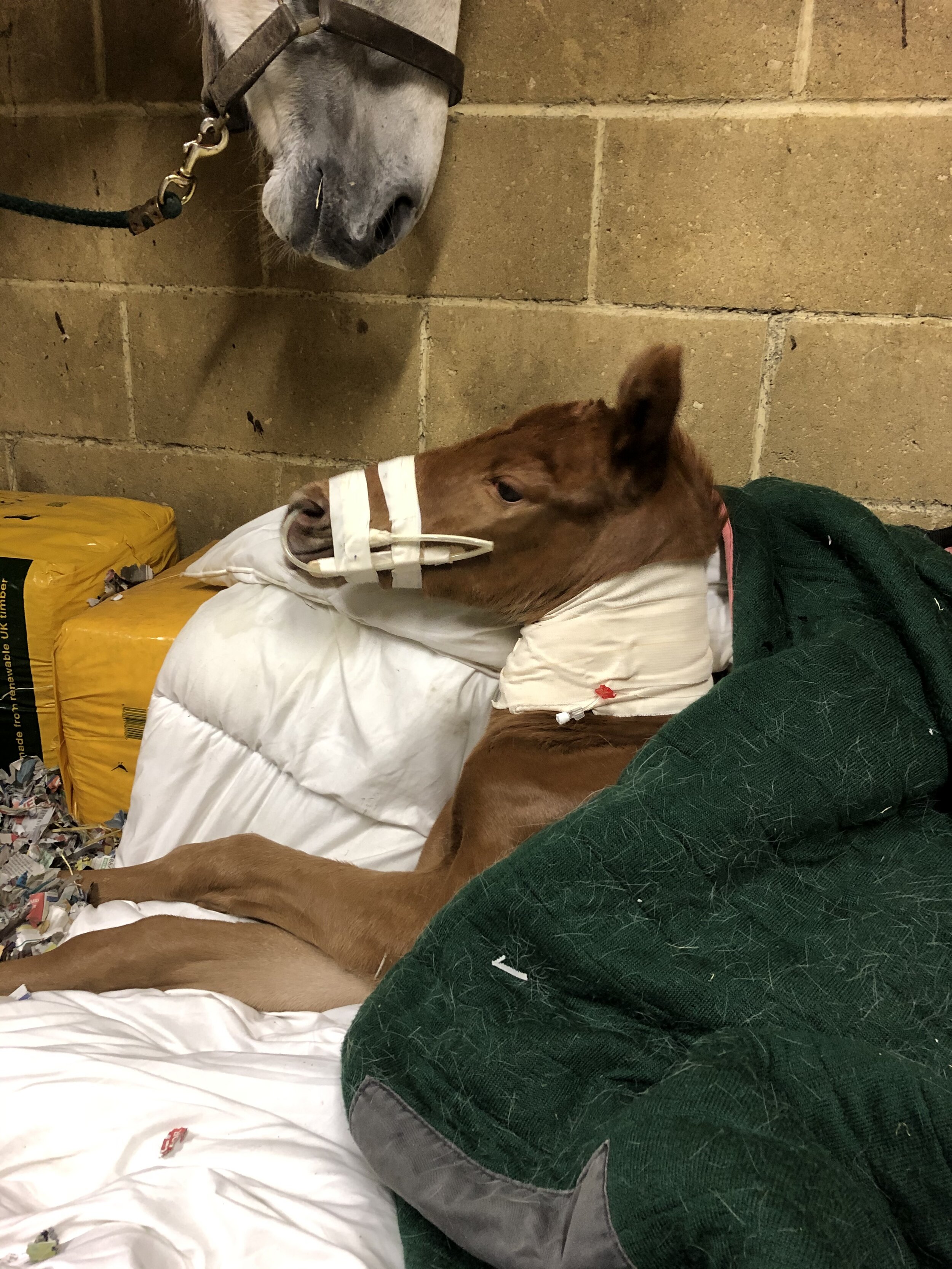
We are very happy to be sending home today, our first critical care foal of the season. He was born two weeks early and suffered from premature placental separation at birth. This in when the placenta comes out with the foal and results in oxygen deprivation or hypoxia. Initially the foal's mucous membranes were a terrible colour due to the lack of oxygen. He was comatose, with no suck reflex . Rae, one of our nurses , administered oxygen for the first hour and his colour started to improve. One of the problems with premature foals is that they haven't produced enough of a chemical called surfactant which is vital to allow full inflation of the lungs. This was clearly an issue with this case. To compound the foal's situation, his mother didn't have a drop of milk so no colostrum was available. Luckily the stud had another mare foal at the same time so we milked off some of her colostrum and fed it to the poorly foal via stomach tube. It was evident that this foal was going to require intensive care if he was to have any chance of survival so he was admitted to the Clinic with his mother. We started her on oxytocin and domperidone to stimulate milk production. The foal was fitted with a feeding tube so that milk could be given easily. We fed him every hour with 200ml of milk replacer, increasing to 300ml by day three. Luckily by day three, the domperidone had stimulated the mare’s milk production so that we could feed the foal the real stuff!

On day two of life the foal started showing the characteristic neurological signs of maladjustment, a delayed response to the oxygen deprivation at birth. This was treated with cortisone therapy and he was given the Madigan foal squeeze procedure which mimics the pressure on the torso normally experienced in the birth canal which is postulated to switch off certain neurosteroids which may keep the foal calm whilst still in the uterus. The intensive nursing was continued around the clock. The foal was also given intravenous fluid therapy to help maintain hydration and he was given two litres of hyperimmune plasma to provide the vital protective antibodies that were not obtained via colostrum. As neonatal foals are very susceptible to infection, broad spectrum intravenous antibiotics were administered throughout the foal's treatment. By day five, the foal showed significant improvement and with help, was able to stand. We held him under the mare and he very quickly started to suckle. By day seven, he was able to get up by himself and get onsuck. We left the feeding tube in place until we were satisfied that he was drinking sufficient milk on his own.

Another problem that we had to deal with was that the foal's urachus which connects the bladder to the umbilicus remained open after birth so that when he went to urinate, urine was coming out of his navel as well as his penis. Luckily we were able to resolve this with conservative treatment in the form of cauterising the naval with silver nitrate and placing a soft rubber ligature around the cord. Sometimes surgery is required to close the urachus.

So a big thank you to all our vets and nursing staff for a fantastic team effort to get this foal through a pretty tough start to life.

Foal with feeding tube in place

Foal with feeding tube in place

All fixed and ready to go home!

All fixed and ready to go home!

Challenging Respiratory Case

As we all know, horses are always picking-up respiratory bugs, both viruses and bacteria. They get a snotty nose, cough for a bit and then get better. We often give antibiotics, sometimes bronchodilators such as clenbuterol ( ventipulmin or dilaterol) and possibly a mucolytic for the extra snotty ones, such as dembrexine (sputolosin ) . If the infection is not clearing up as quickly as we would like then the next step is to do a videoendoscopy allowing us to thoroughly visualise the horse’s airway and to collect samples of mucus which are then examined to determine the cell types present. The sample is also cultured in the lab to see what bacteria we are dealing with and what antibiotics they are sensitive to. The cellular content gives us a good idea on the type of problem we are dealing with and helps to differentiate between an infection and an allergic lung response such as RAO or COPD. All routine stuff, and usually results in a rapid return to a healthy horse.

The case I am going to describe is not particularly sick but has proved very refractory to treatment. He is a national hunt horse that has been retired for four years and has not previously had any respiratory problems.

He developed a cough and a snotty nose so he was prescribed with a five day course of oral potentiated sulphonamide antibiotics ( Trimediazine ) and given plenty of turnout to assist with lung drainage. After two weeks there was no improvement. His lungs sounded pretty normal to listen to and he didn’t have a temperature. His blood picture was unremarkable with a normal white cell count and differential. We then endoscoped the horse; the upper airway was normal, no inflammatory response in the throat and both guttural pouches clean. In the lower airway, the trachea, there was a small pool of mucus which didn’t look particularly exciting but we sampled it anyway. To our surprise the cytology of the sample showed an extreme density of white cells both acute inflammatory neutrophils and an the much larger macrophages , essentially it was pure pus! The sample was submitted for culture but unhelpfully, no bacteria grew. This is quite common with such a high density of white blood cells because they literally kill everything locally but clearly were not capable of totally resolving the problem. So without the help of a positive culture to direct us towards the appropriate antibiotic we chose doxycycline ( Karadox ) which is a broad spectrum drug with good tissue penetration and is typically good at killing pathogenic respiratory bacteria. As there is often an allergic component of the inflammatory response in the equine lung, we opted to give the horse some corticosteroid ( beclamethasone ) by inhaler to try and reduce the degree of inflammation. We also incorporated dembrexine in the treatment plan to loosen up the residual mucus in the lung to help it’s clearance. The horse was given this cocktail of drugs for 10 days and then returned to the Clinic for a repeat endoscopy. As this was billed as a “challenging case” you will not be surprised to learn that the horse had not improved, in fact there was more mucus visible and it was just as pus-like as before in terms of dense cellular content. This case was clearly not going to plan and not responding to treatment so we decided to phone a friend or friends in the form of Prof. Celia Marr from Rossdales and Dr Tim Brazil from Equine Medicine on the move , both world renowned experts in Equine Medicine. They were both very interested in the case and both cited similar cases which had turned out to have long standing walled-off abscesses in the chest. They suggested that we should obtain images of the lungs either via an ultrasound scan or by radiography in order to rule out an abscess. Since we have recently installed a new state of the art HD radiography system at Peasebrook, we were keen to see what quality images we could obtain. As you will see from the picture below the clarity of the lung is exceptional and thankfully it does not show the presence of an abscess.

00000000_DICOM_DX_2019_12_10_0000010512.jpg

So in the light of the radiography findings, both Tim and Celia felt that we must be dealing with an extreme allergic response from the horse, despite the absence of the typical lung sounds normally present in these cases. The recommended treatment is a stronger administration of corticosteroids via the oral route for 14 days plus a strict dust free environment and plenty of turnout. This treatment is ongoing and we will report on the horse’s progress in due course. Fingers crossed that his immune system starts to cooperate with us! This case highlights that it is always best practice to scope and collect samples from horses with respiratory disease if it is not resolving quickly.

Arthroscopic treatment of a flexor tendon injury

This case involves a ten year old hunter mare who presented with a sudden onset forelimb lameness whilst in light work and having had no history of any traumatic episodes. The most salient feature of the clinical examination was distension of the digital tendon sheath or windgall formation and absence of any foot pain. After a thorough aseptic preparation, we blocked the tendon sheath with local anaesthetic; this procedure abolished the lameness. The following day, once all the local had dispersed, we carried out an ultrasound examination of the soft tissue components within the tendon sheath. We also radiographed the fetlock joint to rule out any fractures to the sesamoid bones. Both types of imaging failed to reveal any significant damage to explain the lameness. We therefore decided to medicate the tendon sheath with short-acting cortisone in case the problem was just a synovitis or inflammatory response to trauma. This had the effect of completely reducing the windgall and the mare was rendered free from lameness. She was therefore put back into light walking exercise. Unfortunately the tendon sheath effusion and the lameness soon returned indicating that there had to be an injury which we had not yet identified. The next step with cases displaying this type of history, is to perform an arthroscopic examination of the tendon sheath under general anaesthesia. We took the mare to surgery and we found the problem; there was a substantial split in the medial border of the superficial digital flexor tendon within the annular ligament with a small flap of tissue hinged off the tear. In addition, there was tearing of the medial border of the deep digital flexor tendon. The torn flap of tendon was excised and the remaining strands of tissue were removed using a motorised ressector.

Debridement of the tear in the superficial flexor tendon .

Debridement of the tear in the superficial flexor tendon .

So the free fibres of damaged of tendon were leading to a synovitis or inflammation of the tendon sheath resulting in lameness. Debriding or trimming of all this tendon tissue will allow the inflammation to settle and for the tendon to heal. We will review the comfort level of the horse six weeks post surgery and re-medicate the sheath now that the instigating cause has been removed. All being well, the mare will resume walking exercise at 8 to 10 weeks post surgery. She will stay in a controlled walking regime for 6 to 8 weeks prior to paddock rest for a further 6 to 8 months. The cause of the injury remains a mystery, but must have resulted from some paddock craziness!!

The white structure in the nine o’clock position is the free flap of tendon. Note the inflamed tissue in the background indicative of synovitis.

The white structure in the nine o’clock position is the free flap of tendon. Note the inflamed tissue in the background indicative of synovitis.

Hindlimb Proximal Suspensory Desmitis Update

Hind limb proximal suspensory desmitis (PSD) is a common condition in sports horses and usually presents in one of three ways:

  • unilateral hindlimb lameness

  • bilateral hindlimb

  • no clear lameness but horse displaying rideability problems

PSD describes inflammation and / or damage to the upper region of the suspensory ligament at the top of the cannon bone where the structure mostly originates. Accurate diagnosis is challenging and requires careful interpretation of regional nerve blocking procedures, high quality ultrasound images of both the weight-bearing and non weight-bearing limb. Radiography should be carried-out to rule out co-existing bone pathology. Interestingly, low-field MRI which is the technology used in all the standing MRI units, is not very reliable for identifying suspensory pathology but is useful for picking-up more subtle bone lesions in the upper cannon bone region. Ultrasound scans can show enlargement of the proximal suspensory ligament, loss of fibre pattern and adhesions to surrounding structures.

Conservative treatment of PSD involving rest, medication and remedial shoeing is often not very effective.Unfortunately, surgery which entails removing a section of the deep branch of the plantar nerve and releasing the fascia surrounding the suspensory ligament is the only reliable treatment option giving a 70-80% chance of resolving the lameness providing any additional problems are also addressed; for instance around 40% of PSD cases are also found to have sacro-iliac pain. This essentially denerving surgery, should ideally only be performed on cases where there is minimal fibre disruption so that the structure of the ligament does not fail when it is subsequently, fully loaded post-surgery. This is particularly true for horses with a large hock angle i.e. those with a rather straight hind leg. Studies have shown that a hock angle of 165 degrees or higher is associated with an increased incidence of PSD. So next time you are contemplating a new purchase, take a protractor with you!

This large hock angle conformation predisposes to PSD

This large hock angle conformation predisposes to PSD

Equine Flu Vaccination Update

Since the equine influenza outbreak in February this year, the vast majority of horse owners have wisely had their horses vaccinated or have had boosters to raise antibody levels. As you will be aware many organisations and societies, including the BHA who run Britain’s horse racing, have insisted that horses entering licensed premises or competition venues have had a flu booster within the previous 6 months, assuming they have already had their primary course. So where do we go from here? We thought it would be helpful provide a summary of what is required by the various equine disciplines going forward;

Thoroughbred Racing: As from 17th September 2019, the BHA has relaxed requirements for all horses entering racecourses including runners from non-licensed yards e.g. hunterchasers, nonGB runners (including Ireland and France) and other horses such as ROR competitorsand pony racers. From this date, horses must have had a booster within the previous nine calender months (ideally within 8 months but they are allowing one months grace just to make things more complicated!). This 8 month booster rule applies once the horse has had a primary course of two injections given between 21 and 92 days apart followed by a third injection given between 150 and 215 days after the second injection. Horses are considered to be vaccinated and therefore are entitled to race, after they have had their first two injections but have not yet had the third. The BHA veterinary committee will continue to monitor the prevalence of flu and could change these new requirements if they deemed necessary.

British Eventing: No Horse may take part in a BE National Event (which includes entering competition stables) unless it has a current vaccination against equine influenza which complies with the following conditions:

  • Two injections for primary vaccination, not less than 21 days and not more than 92 days apart, are required before being eligible to compete;

  • A first booster injection must be given within seven months after the second injection of primary vaccination;

  • Subsequent booster injections must be given at intervals of not more than one year, commencing after the first booster injection;

  • The most recent booster injection must have been given within the six calendar months prior to the horse arriving at the competition.

Forgotten passport. Any horse without a passport will be sent home (plus travel companions).

Unvaccinated companion horse. Passports and vaccination records in accordance with the new rules must be carried for all horses on board any vehicle. Any horses without passports and compliant vaccination records will be asked to leave the site, along with any others which they may have travelled with.

Booster given within seven days of the Event. This is fine. As long as the vaccination was given at least the day before the horse arrives at the Event, and is not more than one year after the previous booster. (see Rule 10.2.4)

Only primary course given. This is fine, as long as the horse has had the first two injections that make up the primary course, and the second injection was given within the last six months.  

Primary course given, but first booster (due within seven months) has not yet been given. Fine if second injection was less than six months ago.

Historical discrepancies (ref: rule 10.2.3). In cases where there are historical discrepancies (e.g. booster was given five days late in 2014), but the primary course is correct and the horse has had the most recent booster within the last six months, it will be at the discretion of the Vet and BE Steward as to whether the horse may compete.

British Show Jumping: Flu vaccinations are mandatory for all registered horses and ponies and they must be in possession of a valid flu vaccination certificate. It is the owner's responsibility to ensure that the horse's vaccinations are up to date and correctly recorded on the diagrammatic vaccination record. Spot checks will be regularly carried out at shows. The horse/pony must have received two injections for primary vaccination against equine influenza given no less than 21 days and no more than 92 days apart. Only these two injections need to have been given before a horse/pony can compete in competitions. In addition, a first booster injection must be given no less than 150 days and no more than 215 days after the second injection of the primary vaccination. Subsequently, booster injections should be given at intervals not more than a year apart.

British Dressage: Any horses competing under BD rules at any level must be fully vaccinated.Rule 9 (p58 of the 2019 Members’ Handbook) states: To protect the health of the other competing horses and the biosecurity of the venue, a valid passport must accompany the horse to all competitions and be produced on request. Failure to comply is a disciplinary offence and will debar the horse from competing at the event for which it has been entered. A horse will not be permitted to compete unless it has a current vaccination against equine influenza which complies with the following conditions:
- An initial course of two injections for primary vaccination, not less than 21 days and not more than 92 days apart, are required before being eligible to compete
- A first booster injection must be given between 150 and 215 days after the second injection of primary vaccination
- Subsequent booster injections must be given at intervals of not more than one calendar year, commencing after the first booster injection
- The full course or booster must have been administered at least seven days before the competition.
The vaccination record(s) in the horse’s passport, must be completed, signed and stamped line by line, by an appropriate veterinary surgeon (who is neither the owner nor the rider of the horse).

The 2020 Members’ Handbook will mandate six-monthly Equine Influenza boosters, instead of yearly vaccinations. If you’re currently outside the six month period from your last vaccination, we recommend you have a booster, but you may wait until your next annual renewal date to start your six-monthly vaccination. There’s no need to restart with an initial course, unless otherwise advised by your vet.

The requirement of 'the full course or booster must have been administered at least seven days before the competition' remains the same as in previous years.

Any horse found without adequate and up to date vaccinations will not be allowed to compete with BD and will be suspended until this is rectified.

FEI : All proprietary Equine Influenza vaccines are accepted by the FEI, provided the route of administration complies with the manufacturer’s instructions. An initial Primary Course of two vaccinations must be given; the second vaccination must be administered within 21-92 days of the first vaccination. The first booster must be administered within 7 calendar months following the date of administration of the second vaccination of the Primary Course. Booster vaccinations must be administered at a maximum of 12 month intervals however horses competing in Events must have received a booster within 6 months +21 days (and not within 7 days) before arrival at the Event. Horses may compete 7 days after receiving the second vaccination of the primary course. Horses that have received the Primary Course prior to 1 January 2005 are not required to fulfil the requirement for the first booster ( 7 month ), providing there has not been an interval of more than 12 months between each of their subsequent annual booster vaccinations.

Pony Club: A valid passport and vaccination record:

must accompany the horse/pony to all events

must be available for inspection by the event officials

must be produced on request at any other time during the event .

Subject to paragraph * below, no horse/pony may take part in an event (which includes entering competition stables) unless it has a Record of Vaccination against equine influenza which complies with the Minimum Vaccination Requirements.

The Minimum Vaccination Requirements for a horse/pony are:

(a) if the current vaccination programme started BEFORE 1 January 2014 that it has received:

a Primary Vaccination followed by a Secondary Vaccination given not less than 21 days and not more than 92 days after the Primary Vaccination; and

if sufficient time has elapsed, a booster vaccination given not less than 150 days and not more than 215 days after the Secondary Vaccination and further booster vaccinations at intervals of not more than a year apart

PROVIDED THAT if all annual boosters given AFTER 31 December 2013 have been given correctly, any error with the first booster vaccination or an annual booster given BEFORE 1 January 2014 may be ignored

(b) if the current vaccination programme started AFTER 31 December 2013 that it has received:

• a Primary Vaccination followed by a Secondary Vaccination given not less than 21 days and not more than 92 days after the Primary Injection; and

• if sufficient time has elapsed, a booster vaccination given not less than 150 days and not more than 215 days after the Secondary Vaccination and further booster vaccinations at intervals of not more than a year apart.

The Record of Vaccination in the pony’s passport must be completed by a veterinary surgeon, signed and stamped line by line

No horse/pony whose latest booster vaccination is more than 14 days overdue may take part in a competition under any circumstances.

*Notwithstanding the above in cases where the Event Veterinary Officer, following consultation with the Pony Club Steward, is satisfied that the presence of the horse/pony at the event does not pose a threat to bio- security at the event, that horse/pony may nonetheless take part in the event on such conditions as the Event Veterinary Officer considers appropriate, but the circumstances must be noted on the certificate. Any horse/pony allowed to compete under this discretion must be re-vaccinated to comply with the Minimum Vaccination Requirements and the certificate duly completed before it is eligible to compete again.

No pony may compete on the same day as any relevant vaccination is given or on any of the 6 days following such a vaccination.

Clearly, many Pony Club activities take place at venues used for other equine disciplines and therefore such venues may have in place, more stringent rules regarding Equine Influenza vaccination. Many local venues are requesting that horses and ponies must have had a booster within the previous 6 months prior to competing and often that injections must not have been administered during the previous 7 days. Racecourses now require that boosters must have been given within the previous 9 months, so there is lots of scope for confusion. If in doubt and you use many different venues, it is prudent to adopt the 6 month booster strategy to avoid problems.

British Riding Clubs: This rule applies in respect of any horse or pony which competes in a BRC Area Qualifier and Championship. Section 2 20 BRC MEMBERS HANDBOOK The horse or pony must have been vaccinated against equine influenza by a veterinary surgeon who is not the owner of the animal, in accordance with the following rules: The horse or pony must have received a primary injection followed by:

• a second primary injection which is given not less than 21 days and not more than 92 days after the first

• a first booster injection which is given not less than 150 days and not more than 215 days after the second primary injection

• further annual booster injections at intervals of not more than a year apart.

If the current vaccination programme started AFTER 1 January 2014:

• the first two primary injections must be correct i.e. the second given between 21 and 92 days after the first

• the first booster must be given between 150 and 215 days after the second primary injection

• all annual boosters must be correct. However, any errors with first booster (which should be given 150 – 215 days after the second primary injection) or annual booster given BEFORE 1 January 2014 may be ignored provided that:

• the first two primary injections are correct i.e. the second given between 21 and 92 days after the first

• all annual boosters given AFTER 1 January 2014 are correct. Leap years will be ignored for an annual booster, but for the two primary injections and first booster injection, the days must be counted and therefore a leap year would interfere with the correct number of days between injections.

Horses may compete at BRC Competitions providing that they have had the first two primary injections. No injection should have been given on any of the 6 days before a competition or entry to championship stables. For example: if the horse is vaccinated on the Monday, the horse will not be eligible to enter championship stables, or compete until the following Monday.

In the event of failure to comply with any of the requirements of this rule, the horse or pony will be disqualified and not permitted to take part in any competition to which these rules apply.

Checking of Passports and Equine Influenza Records; horses must be presented in a bridle to the flu vac checker at Championships and where applicable Area Qualifiers. For the purposes of determining whether the requirements of these rules have been met, the following documents must be available for inspection in respect of a horse or pony which is taking part in a BRC Area Qualifier or Championship.

• any passport issued for the horse and

• the full vaccination records for the horse if this is not contained in the passport

The identification of the horse or pony must be checked against that contained in the passport or on the flu vaccination record. This may be done from the diagram and description of the animal or by microchip providing that the microchip number has been recorded in the passport or flu vaccination record.
Nail penetration of the sole

Recently we have had a couple of cases of nail penetration of the foot which highlighted the potential risks of this injury and the need for immediate correct treatment. Excellent knowledge of the anatomy of the foot is crucial to understand where a penetrating object can reach and why the consequences can be so serious.

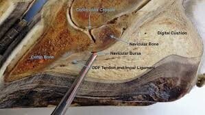
Sagittal section through the hoof, the pointer is marking the position of the navicular bursa.

Sagittal section through the hoof, the pointer is marking the position of the navicular bursa.

As you can see from the image above, a nail penetration in the direction of the pointer will pass through the digital cushion, the deep flexor tendon and into the navicular bursa. Inevitably the nail will introduce dirt and bacteria into these sensitive areas. If navicular bursal penetration is not treated immediately, the structure will become chronically infected and ultimately the horse would have to be euthanased on welfare grounds. So accurate evaluation of the injury is vital to ensure that the correct treatment options are employed. If possible, we take x-ray images of the foot with the nail in situ so that we can appreciate its trajectory. Often, however, owners have already removed the nail prior to calling the vet. In these cases, after thoroughly cleaning the sole, we attempt to introduce a metal probe into the hole left by the nail so that we can see that on our x-ray instead. We can also inject a radio-opaque dye into the hole which shows up on x-rays and helps us assess the extent of the penetration. MRI is another option, if readily available, to evaluate which structures have been compromised

Probe inserted into nail penetration tract .

Probe inserted into nail penetration tract .

The radiographic image above is from one of our recent cases; it shows the nail will have penetrated through the digital cushion and the deep flexor tendon and will therefore have introduced bacteria close to the navicular bursa.This horse was extremely lame. The foot was prepared with a rasp and hoof knife so that all loose tissue was removed. The whole foot was then dressed in iodine-soaked bandages for an hour prior to being taken to surgery. Under general anaesthesia the penetrating tract through the frog was explored and enlarged so that the digital cushion and deep flexor tendon could be flushed with sterile saline under high pressure. The arthroscope was then introduced via a portal in the pastern region, down into the navicular bursa. This structure was explored for signs of penetration, which fortunately had not occurred in this case, and was then flushed using an exit portal. Antibiotics were then deposited into the bursa. If there had been penetration of the bursa, then flush fluids would have found there way out through the frog wound, this obviously didn’t occur in this case. At the end of the surgical procedure the foot and the lower limb were dressed very carefully with sterile protective dressings. These cases are put on high doses of strong broad-spectrum antibiotics for a minimum of five days. With this case the recovery has been uneventful and following two weeks of dressings, the foot was then fitted with a shoe and hospital plate which allows us access to the frog region to regularly inspect the healing frog wound.

Shoe fitted with a hospital plate to allow access to the healing penetration wound in the frog.

Shoe fitted with a hospital plate to allow access to the healing penetration wound in the frog.

The second of the recent cases involved a nail penetration in the cleft of the frog just in front of the site of attachment of the deep flexor tendon to the pedal bone. The nail had been removed by the owner and so after thorough cleansing, a probe was introduced into the hole to establish the trajectory . Luckily although it went down to bone, it was heading forwards and so missed the tendon, but only just! With this case, the hole was opened-up to aid flushing and to provide a drainage route for any pus. The foot was poulticed for two days then dressed with iodine soaked bandages. This horse was put on the same broad-spectrum combination of antibiotics and no pain relief was allowed so that the horse’s true level of comfort was known. His recovery was straightforward. It is absolutely crucial to make sure that these patients have been vaccinated against tetanus as they would be nailed-on (excuse the pun) to get it otherwise. If in doubt, tetanus antitoxin should be given to provide immediate cover.

So in summary, if your horse is unlucky enough to suffer from a nail penetration of the foot, call us straight away and try and resist the temptation to remove the nail!

Calcaneal Bursoscopy

On the 10th of July this year, a 10 year old brood mare was referred to us by another practice for swelling and heat in the calcaneal bursa area of the right hind (at the back of the hock). She was very lame (4/5) and painful on palpation of that area. There had been history of a tiny wound on the inside of the hock but when she came in, there was no evidence of open wounds. 

When she arrived, the mare was sedated for a thorough ultrasound scan of the hock area, which showed evidence of severe inflammation of the sub-tendineous calcaneal bursa. This bursa is located between the superficial flexor tendon, the gastrocnemius muscle and the calcaneus, it prevents the tendon from rubbing against the bone surface.

This is a pathology that should be taken very seriously, especially in the chronic stage. The prognosis is guarded and aggressive treatment is required in order to acquire healing. We decided to perform bursoscopy in order to flush the calcaneal bursa so that the inflammation could settle and the pressure would be taken off the surrounding tissues. We also performed IVRA (intravenous regional antibiosis) in the affected limb during surgery. In order to do this, a tourniquet was placed above the hock and antibiotic was injected into the metatarsal vein. By leaving the tourniquet in place for 10-15 minutes, the antibiotic can diffuse locally and obtain high concentrations at the infection site.

The surgery went very well and the mare is sound after about a month of recovery despite the poor prognosis.

On this ultrasound image distension of the bursa is visible, containing strands of fibrin

On this ultrasound image distension of the bursa is visible, containing strands of fibrin

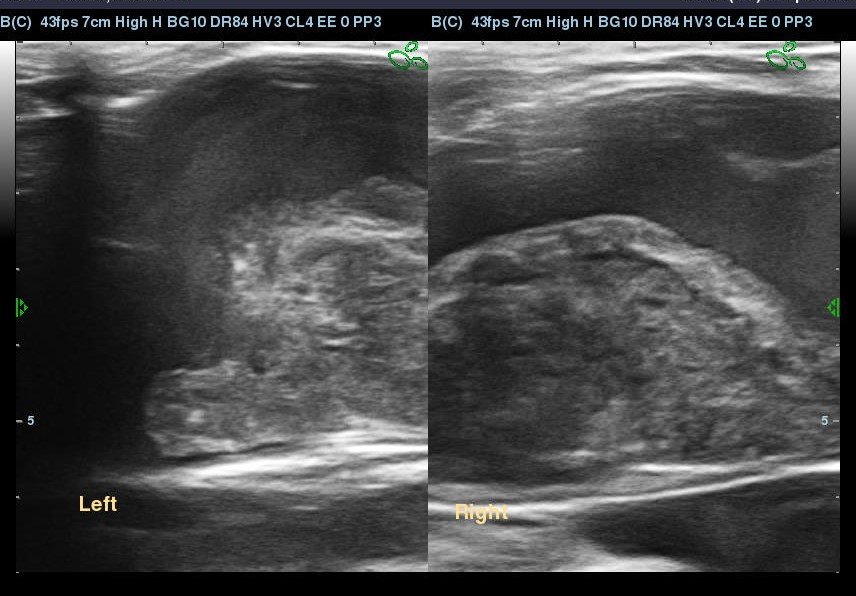
On this image you can see the difference between the affected bursa (right hind) and the normal limb (left hind). On the image of the right hind the superficial flexor tendon is visible at the top of the image and the gastrocnemius tendon is visible…

On this image you can see the difference between the affected bursa (right hind) and the normal limb (left hind). On the image of the right hind the superficial flexor tendon is visible at the top of the image and the gastrocnemius tendon is visible at the bottom, separated by the droplet shaped inflamed bursa.

Bursoscopy image from during the surgery looking inside the calcaneal bursa. The red fibrous aspect of the inside of the bursa is an indication of severe inflammation.

Bursoscopy image from during the surgery looking inside the calcaneal bursa. The red fibrous aspect of the inside of the bursa is an indication of severe inflammation.

Bursoscopy image where you can see the fibrous/flaky content of the inflamed bursa. This was debrided and flushed out.

Bursoscopy image where you can see the fibrous/flaky content of the inflamed bursa. This was debrided and flushed out.

Anatomy of the hock of the horse. “calcanean bursa” indicates the subtendineous bursa we performed surgery on (situated between the superficial flexor tendon and the gastrocnemius muscle.

Anatomy of the hock of the horse. “calcanean bursa” indicates the subtendineous bursa we performed surgery on (situated between the superficial flexor tendon and the gastrocnemius muscle.

Gastroscopy Follow-up

On July 9th, we published some images relating to a patient that had been diagnosed with both squamous and glandular stomach ulcers but had not shown any clinical signs whatsoever, highlighting the policy of routine gastric screening in horses. Untreated ulcers, as well as being extremely painful , can lead to perforation of the stomach wall , peritonitis and death, so we take them very seriously!

Thankfully the horse mentioned in the previous blog responded well to treatment with omeprazole (Peptizole) to reduce gastric acid production, coupled with sulcralfate which coats the stomach wall to protect the damaged tissue. He was re-scoped yesterday and the images are seen below:

This image shows the junction of the squamous and glandular regions, now with no ulceration.

This image shows the junction of the squamous and glandular regions, now with no ulceration.

IMG0002.jpg

As can be seen, this horse has responded well to our normal treatment regime but not all patients do so well. A recent review article in Equine Veterinary Education by B W Sykes, highlighted the fact that around 15 to 30% of squamous cases and up to 75% of glandular ulceration patients fail to respond to normal treatment. It is postulated that there may be marked variation in individual’s uptake of omeprazole and this can be affected by the timing of drug administration and feeding strategies. For instance, we advise generally that ulcer patients have ad lib fibre but Sykes suggests that omeprazole uptake will be more optimal if given after a period of fasting i.e. first thing in the morning when hopefully the horse finished his late in the evening and also around 60-90 minutes before giving sulcralfate which in coating the stomach wall, will affect omeprazole uptake. The slow-release intramuscular omeprazole injection should definitely be considered if oral dosing is proving ineffective. Work is also being done to evaluate an alternative proton pump inhibitor to omeprazole, called esomeprazole. Studies have shown that this new drug produces more pronounced suppression of gastric acid at lower dose rates than omeprazole.

The importance of Gastroscopy

Two weeks ago we discovered stomach ulcers in one of our patients by performing a gastroscopy. Interestingly the horse did not show specific signs of gastric ulceration. We decided to perform a routine gastroscopy since the horse recently moved yards and underwent a stressful journey. To our surprise we discovered quite severe glandular  (grade 3/4) and non-glandular (grade 2/4) ulceration (shown in the videos beneath).

==> in this video you can see the clear separation between the glandular part of the stomach (pink) and the non-glandular part (white/beige). The separation between the two parts is called the “margo plicatus” and this particular region is very prone to ulceration. In this horse you can see bleeding ulcers along the margo plicatus.

==> in this video you can see the “Pylorus”, this is the exit of the stomach towards the small intestine and is part of the glandular part of the stomach. Glandular ulceration is visible all around this exit (orange coloured).


Gastric ulcers can either cause obvious clinical signs such as: reduced or selective appetite,lethargy,  being sensitive to girth up, losing weight, colic (scraping, flank watching/biting) or yawning. Unfortunately the signs can also be very subtile such as: being of colour, losing condition, dullness of the coat, behavioural problems and more.

This condition is quite easily treatable with Omeprazole (for squamous ulcers) and Sucralfate (for glandular ulcers) for usually at least a month. Another important factor is the management of the horse. Reducing stress, giving multiple meals a day, ad libitum hay/haylage and a feed high in protein (13-14 %) are the main changes you can apply.

The horse will be re-scoped in a month's time to check whether the treatment has been success. An update will follow with the results.

Foal Wound and skin graft

A Progress update from the foal who tried to jump a fence.

The wounds remained closed for a few weeks, this was vitally important to keep to exposed bone covered and prevent sequester formation. Sequestration is where an area of bone dies and becomes like a foreign body in need of surgical removal.

The skin around the wound began to die back and the wound contracted - this is a normal phase of wound healing. Over the next month the foal underwent regular bandage changes. The wound took time to fill in and when it did there was exuberant granulation tissue - proud flesh.

Proud flesh is common in equine wounds, especially on the distal limb. It is treated by excising the tissue that is above (proud) to the skin - thus allowing the skin to epithelialise over the top.

In this case as the wound was so large, the decision was taken to take skin grafts from the neck and place them in the wound. These islands of skin promote skin growth over the top of the wound.

As you can see from the photos the wound is closing up well and in a few weeks we should have a completely healed leg.

As you can see from this last photo, there is a different texture to the tissue between the islands of graft - this is skin beginning to heal over the top

IMG_0048.JPG